

Beyond the Waveform: How AI Is Expanding the Diagnostic Potential of the 12-Lead ECG
How deep learning is transforming the 12-lead ECG into a high-dimensional tool for detecting structural and functional cardiac phenotypes.
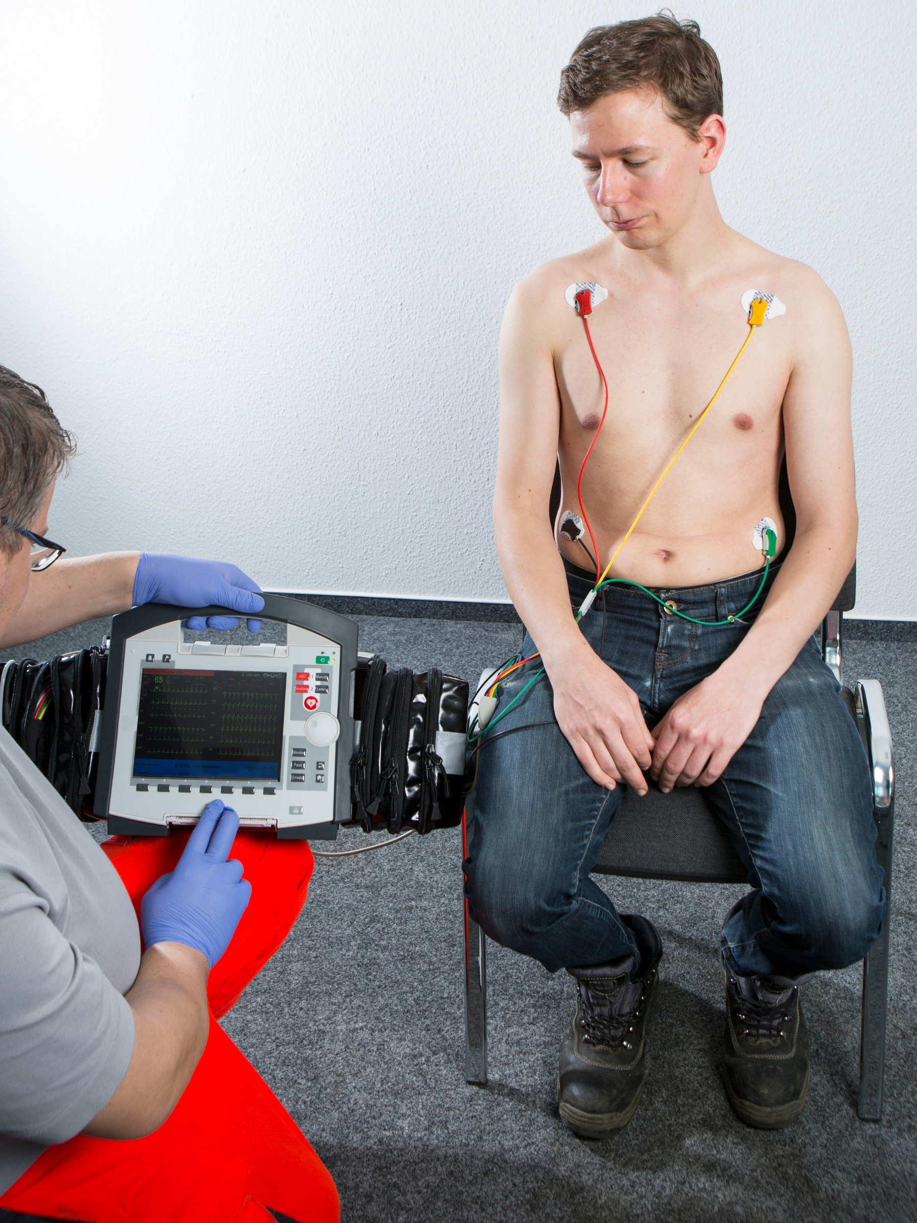
A routine 10-second ECG contains tens of thousands of digital measurements. Deep learning is beginning to demonstrate that those signals may encode clinically meaningful information about cardiac structure and function that extends beyond conventional electrocardiographic interpretation.
For more than a century, the 12-lead electrocardiogram has been foundational to cardiovascular medicine.
Its traditional diagnostic utility is well established: rhythm and conduction abnormalities, repolarization disturbances, chamber abnormalities, ischemic changes, infarction patterns, and numerous other electrophysiologic findings.
What is changing is not the ECG acquisition itself.
It is the analytical depth that can be applied to the underlying signal.
Artificial intelligence—particularly deep learning—allows the raw digital ECG waveform to be interrogated at a dimensionality that conventional visual interpretation cannot practically reproduce.
Rather than asking only whether an ECG demonstrates a recognized abnormality, AI-enabled electrocardiography introduces a different question:
Can the electrical phenotype contain reproducible signatures of structural or functional cardiovascular disease that are not readily apparent through conventional ECG interpretation?
An expanding body of research suggests that, for certain phenotypes, it can.
That possibility could fundamentally broaden the role of one of medicine's oldest diagnostic technologies.
The ECG as a High-Dimensional Physiologic Dataset
The familiar ECG tracing represents only the visual expression of a much larger numerical dataset.
Medical AI's AiTiA platform analyzes the raw digital ECG signal rather than relying solely on an image of the tracing.
At a sampling frequency of 500 Hz, each lead generates 500 numerical measurements per second.
Across a standard 10-second, 12-lead acquisition:
500 samples/second × 10 seconds × 12 leads = 60,000 numerical data points.
Medical AI's technical materials specifically identify these 60,000 raw measurements as the input available for AI analysis.
Conventional interpretation reduces this information into clinically meaningful constructs: rhythm, rate, intervals, axis, voltage, morphology, conduction, depolarization, repolarization, and recognized patterns of disease.
Deep-learning models are not necessarily constrained to those predefined features.
They can learn latent representations within the raw waveform—complex combinations of temporal, morphologic, amplitude, and interlead relationships statistically associated with a defined clinical phenotype.
Some of these representations may overlap with electrocardiographic features clinicians already recognize.
Others may be distributed across the waveform and not readily reducible to conventional human-interpretable ECG criteria.
This is the fundamental premise underlying AI-ECG.
Connecting the Electrical Phenotype to the Echocardiographic Phenotype
Left ventricular systolic dysfunction provides an instructive example.
An ECG does not directly measure ejection fraction.
Echocardiography does.
For an AI-ECG model targeting LV systolic dysfunction, ECG recordings can be paired with a reference phenotype established by cardiac imaging.
The model is then trained to identify features within the electrical signal associated with the imaging-defined phenotype.
In simplified terms:
Echocardiography establishes the cardiac phenotype.
The ECG provides the electrical phenotype.
Deep learning identifies statistical relationships between the two.
For AiTiA LVSD, Medical AI defines the target phenotype as LVEF ≤40%.
The algorithm therefore does not "calculate" LVEF from an ECG in the manner that an echocardiographic measurement is obtained.
Nor does it create a surrogate echocardiographic image.
It identifies an ECG-derived probability signal associated with the presence of an echo-defined ventricular phenotype.
That distinction is critical to interpreting both the capabilities and limitations of the technology.
Why Should LV Dysfunction Produce an AI-Detectable Electrical Phenotype?
The biological premise is plausible because myocardial structure, ventricular function, and cardiac electrophysiology are interdependent.
Ventricular remodeling, fibrosis, changes in myocardial mass, altered loading conditions, ischemic injury, neurohormonal effects, conduction disturbances, and changes in myocardial function can affect depolarization and repolarization.
Some resulting manifestations are already recognized clinically.
Others may produce far more subtle alterations in waveform morphology, timing, amplitude, and interlead relationships.
A deep-learning architecture can interrogate those relationships simultaneously across the full digital waveform.
The model is therefore not visualizing mechanical dysfunction.
It is detecting a high-dimensional electrical phenotype statistically associated with it.
This distinction is one reason AI-ECG may be better conceptualized as an ECG-derived digital biomarker rather than as an alternative imaging modality.
AiTiA LVSD: Identifying Reduced LV Systolic Function From the 12-Lead ECG
Medical AI's AiTiA LVSD algorithm is designed to identify patients with left ventricular systolic dysfunction using a routine digital 12-lead ECG.
The target condition described in Medical AI's clinical materials is LVEF ≤40%, corresponding to clinically significant reduced LV systolic function.
The acquisition remains a standard 10-second ECG.
The raw waveform is analyzed by the AI model.
The software generates a continuous 0–100 risk score and risk classification that can be incorporated into the existing ECG-reporting workflow.
No additional physiologic acquisition is required simply to generate the AI analysis.
That creates an important distinction between AI-ECG and many other diagnostic technologies.
The diagnostic input remains familiar. The analytical depth changes.
What Does the Clinical Validation Show?
Medical AI's pivotal clinical materials report a single-center study involving 688 participants, with approximately 15% prevalence of LV systolic dysfunction.
Reported performance included:
AUROC: 0.919
Positive predictive value: 0.715
Negative predictive value: 0.982
Medical AI also reports subgroup analyses across age, sex, and medical history, with performance remaining robust within the evaluated subgroups.
In a subgroup for whom NT-proBNP results were available, the company's pivotal-study analysis reported an AUROC of 0.905 (95% CI, 0.842–0.968) for AiTiA LVSD compared with 0.720 (95% CI, 0.635–0.804) for NT-proBNP.
That comparison is potentially interesting, but it should be interpreted cautiously. It represents a smaller subgroup and does not establish AI-ECG as a replacement for natriuretic peptide testing.
More broadly, peer-reviewed evidence in the AI-ECG field supports the feasibility of identifying reduced ventricular systolic function from the 12-lead ECG.
The clinical question, however, extends beyond discrimination.
For physicians considering how such technology could actually be deployed, AUROC is only the beginning.
What the AI-ECG Result Does—and Does Not Mean
A positive AiTiA LVSD result does not directly measure LVEF.
It does not establish a diagnosis of heart failure.
It does not characterize ventricular morphology.
And it does not replace echocardiography when definitive assessment of cardiac structure and function is clinically indicated.
The model generates an ECG-derived risk signal associated with the target phenotype of LVEF ≤40%.
Its interpretation therefore depends on clinical context.
That includes:
- Pretest probability
- Symptoms and functional status
- Known cardiovascular disease
- Comorbidities
- Physical examination
- Prior ECG findings
- Natriuretic peptide data when appropriate
- Previous cardiac imaging
- The prevalence of LV dysfunction in the population being evaluated
- The clinical consequences of false-positive and false-negative results
The prevalence issue is particularly important.
PPV and NPV are not intrinsic characteristics of an algorithm independent of the population in which it is used.
In a low-prevalence screening population, an algorithm can maintain excellent discrimination, sensitivity, and specificity while producing a substantially lower PPV.
In a higher-risk population, the predictive-value profile may be very different.
The clinically meaningful question is therefore not simply:
"What is the AUROC?"
It is:
"Does the algorithm identify the appropriate patients for definitive evaluation, with an acceptable false-positive and false-negative burden, in the population in which it is being deployed?"
That is the standard against which AI-ECG should ultimately be evaluated.
The Appropriate Clinical Role Is Triage, Not Echocardiographic Replacement
At present, the most clinically coherent role for AI-ECG is not as a replacement for echocardiography.
It is as an additional layer of screening, risk stratification, or diagnostic triage.
Echocardiography provides direct structural and functional information that an ECG cannot.
AI-ECG addresses a different problem:
Can a rapidly acquired, widely available physiologic signal identify patients whose probability of ventricular dysfunction is sufficiently elevated to warrant definitive assessment?
Conceptually, the pathway becomes:
Routine 12-lead ECG → AI-derived probability signal → targeted echocardiography or additional evaluation when clinically appropriate.
This distinction has practical significance.
Population-wide echocardiographic screening is resource intensive.
ECGs, by contrast, are already acquired at enormous scale across healthcare.
If AI can extract a validated ventricular dysfunction signal from an ECG that has already been obtained, the technology could create a low-incremental-burden mechanism for identifying patients who might otherwise not undergo imaging.
The objective is not necessarily to perform more echocardiograms.
It is potentially to identify more effectively which patients should receive them.
A Normal-Looking ECG May Not Be Data-Normal
One of the more provocative implications of AI-ECG is that a tracing can appear relatively unremarkable by conventional visual criteria while still contain a disease-associated signal.
Human ECG interpretation relies on recognizable constructs:
Rate.
Rhythm.
PR interval.
QRS duration.
QT interval.
Axis.
Voltage.
Conduction.
ST-T morphology.
Q waves.
Hypertrophy patterns.
An AI model is not restricted to those constructs.
It can evaluate nonlinear interactions distributed across tens of thousands of numerical measurements.
A patient therefore may lack an obvious human-interpretable ECG signature of LV systolic dysfunction while still producing a waveform containing features that contribute to an elevated model output.
This is both scientifically compelling and clinically challenging.
The inability to reduce every model output to a recognizable ECG feature increases the importance of:
External validation.
Calibration.
Population-specific performance.
Prospective evaluation.
Clearly defined intended use.
Workflow integration.
Post-deployment performance monitoring.
The more powerful the inference becomes, the more rigorous the validation must be.
From a Single Algorithm to a Multi-Phenotype AI-ECG Platform
LV systolic dysfunction represents only one potential application.
Medical AI's broader research program describes AI-ECG investigation across multiple cardiovascular and systemic phenotypes, including:
- Left ventricular systolic dysfunction
- Left ventricular diastolic dysfunction
- Valvular heart disease
- Pulmonary hypertension
- STEMI and NSTEMI
- Atrial fibrillation prediction
- Anemia and electrolyte abnormalities
- Cardiac arrest risk
Medical AI's research materials describe published work across this broader spectrum of cardiovascular and systemic conditions.
Its current development pipeline also includes additional disease-specific AI-ECG applications.
This creates an important conceptual distinction.
AiTiA can be viewed not simply as a single LVSD-detection algorithm, but as a potential multi-phenotype AI-ECG platform architecture.
The underlying physiologic input can remain largely unchanged:
a digital 12-lead ECG waveform.
What changes is the independently developed, validated, and appropriately authorized algorithm applied to that waveform.
Additional Indications Could Fundamentally Change the Role of the ECG
Medical AI intends to continue developing additional AI-ECG indications and pursuing the applicable regulatory pathways for clinical use.
Each indication must be evaluated independently.
Performance for LV systolic dysfunction does not establish validity for acute myocardial infarction, aortic stenosis, diastolic dysfunction, pulmonary hypertension, arrhythmia prediction, or another phenotype.
Each clinical application requires its own evidence base, appropriate validation, defined intended-use population, performance characteristics, and applicable regulatory authorization.
But the architecture becomes particularly interesting if multiple disease-specific models ultimately satisfy those requirements.
A single 10-second, 12-lead acquisition could potentially serve as the input for multiple independently validated analytical models.
The same underlying electrical dataset could potentially be interrogated for signatures associated with:
Ventricular systolic function.
Diastolic dysfunction.
Valvular disease.
Myocardial ischemia or injury.
Pulmonary vascular disease.
Future arrhythmic risk.
Other cardiovascular or systemic phenotypes.
The sophistication would not come from making the physical ECG acquisition more complicated.
Quite the opposite.
The acquisition could remain remarkably simple while the computational interpretation becomes increasingly complex.
If multiple independently validated indications ultimately receive appropriate regulatory authorization and can be integrated into a unified clinical platform, AI-ECG could evolve into an unusually sophisticated form of multi-phenotype cardiovascular decision support built on a single physiologic recording.
A Different Model of Diagnostic Technology
Most diagnostic technologies are closely linked to a specific measurement.
Echocardiography generates structural and functional imaging.
Cardiac troponin measures a circulating biomarker associated with myocardial injury.
CT provides cross-sectional anatomical information.
The potential architecture of a multi-indication AI-ECG platform is different.
One physiologic dataset can potentially serve as the input for multiple specialized analytical models.
The physical test does not necessarily have to change when another validated algorithm becomes available.
The analytical layer changes.
This has potentially important implications for clinical scalability.
A healthcare organization does not necessarily need to introduce an entirely new diagnostic acquisition for each AI phenotype.
Instead, additional clinical intelligence may potentially be extracted from a physiologic signal already being collected.
That shifts the paradigm from:
one test → one principal interpretation
toward:
one physiologic signal → multiple independently validated clinical inferences.
That is a fundamentally different way of thinking about the ECG.
Could the AI Score Function as a Longitudinal Digital Biomarker?
Medical AI's research also raises another clinically interesting possibility: whether serial AI-ECG scores may provide information about changes in ventricular function over time.
The company's materials describe cases in which AiTiA LVSD scores increase as ventricular function deteriorates and decrease as function improves.
If validated prospectively for longitudinal use, this could extend AI-ECG beyond binary screening.
Serial ECGs might potentially provide a relatively low-burden digital signal suggesting deterioration or improvement in an underlying ventricular phenotype.
That possibility remains investigational.
It should not currently be interpreted as a replacement for serial echocardiography, clinical assessment, biomarkers, or other established methods of monitoring patients with heart failure.
But the concept is important.
It raises the possibility that the ECG-derived AI score may ultimately function not only as a screening output but as a quantitative digital biomarker whose trajectory contains clinically relevant information.
That hypothesis deserves prospective evaluation.
External Validation and Generalizability Are Critical
High model performance in a development or pivotal cohort does not guarantee equivalent performance across clinical environments.
AI-ECG models can potentially be influenced by differences in:
Patient demographics.
Disease prevalence.
Comorbidity burden.
Referral patterns.
ECG hardware.
Sampling characteristics.
Signal preprocessing.
Clinical setting.
Healthcare-system population.
Disease spectrum.
Threshold selection.
These are not theoretical concerns.
They are fundamental questions for any diagnostic AI technology.
A model intended for broad clinical use should therefore demonstrate that performance remains clinically acceptable outside the population in which it was originally developed.
Medical AI's materials describe international collaborations and external evaluation across multiple institutions and populations.
Continued independent external validation will be particularly important as the technology enters additional healthcare systems and as new indications are developed.
For cardiologists, the question is not simply whether an algorithm works.
It is:
Does it work in my patients, in my clinical environment, at the threshold and prevalence relevant to the decision I am trying to make?
Clinical Utility Must Ultimately Extend Beyond Diagnostic Accuracy
Even excellent diagnostic performance does not automatically establish clinical utility.
A model can discriminate effectively between patients with and without a phenotype while still failing to improve care if its output does not change clinical decisions appropriately.
The critical downstream questions include:
Does AI-ECG identify disease earlier?
Does it appropriately increase confirmatory testing among patients most likely to benefit?
Does it avoid excessive unnecessary imaging?
Does earlier identification result in earlier guideline-directed therapy?
Does it improve referral efficiency?
Does it meaningfully change patient outcomes?
Does it create additional workflow burden?
What is the false-positive burden at scale?
How should clinicians respond to discordance between the AI result and the conventional ECG, symptoms, biomarkers, or prior imaging?
These questions move the discussion from algorithmic performance to clinical utility.
That is where the ultimate value of AI-ECG will be determined.
Workflow Integration Is Part of the Clinical Intervention
Medical AI's proposed implementation integrates AI analysis with the existing ECG and hospital-information workflow.
In its technical materials, the AI-ECG order is entered through the hospital information system, the ECG is acquired, and the resulting AI score and interpretation are incorporated into the existing ECG report.
This matters.
Physicians do not need another isolated portal generating another stream of alerts.
The AI output should appear where clinicians already interpret cardiovascular information and should connect to a defined clinical response.
A positive result must answer the practical question:
What should I do next?
Depending on the indication, patient population, regulatory labeling, and clinical circumstances, that could include:
- Review of symptoms and examination findings
- Comparison with previous ECGs
- Natriuretic peptide testing
- Echocardiography
- Review of prior cardiac imaging
- Cardiology referral
- Additional disease-specific evaluation
AI without an actionable pathway risks becoming another data element.
AI embedded into an evidence-based clinical workflow has the potential to become decision support.
Opportunistic Screening May Be One of the Most Important Applications
ECGs are already acquired across a wide range of clinical environments:
- Primary care
- Emergency medicine
- Preoperative evaluation
- Cardiology
- Oncology
- Chronic disease management
- Hospital admission
- Routine health evaluation
Many patients undergo ECGs for reasons unrelated to screening for LV systolic dysfunction.
If an appropriately validated and authorized algorithm can extract another clinically meaningful signal from the same digital recording, an ECG already being performed becomes an additional opportunity for disease detection.
This is the concept of opportunistic screening.
Instead of asking:
"Should we perform another screening procedure on every patient?"
AI allows us to ask:
"What additional validated clinical information can be extracted from physiologic data we are already collecting?"
For healthcare systems performing large volumes of ECGs, that distinction could become significant.
AI Does Not Need to Replace the Cardiologist to Transform the ECG
The relevant benchmark is not whether artificial intelligence can replace a cardiologist.
It should not.
The more meaningful question is whether AI can extract reproducible information from physiologic data that helps physicians:
recognize disease earlier,
identify patients for definitive testing more effectively,
quantify previously inaccessible risk signals,
or
identify clinically relevant phenotypes that conventional ECG interpretation may not reveal.
AI-ECG is particularly compelling because it applies that concept to one of the oldest, fastest, and most ubiquitous diagnostic tests in cardiovascular medicine.
The traditional ECG converts cardiac electrical activity into a waveform physicians can interpret.
AI introduces an additional analytical layer.
It interrogates the underlying digital signal at a dimensionality that human visual interpretation cannot practically reproduce.
The result is not a replacement for physician judgment.
It is potentially a new class of digital biomarker derived from a familiar physiologic test.
The Future of the 10-Second ECG
The same 10-second recording can continue to provide the rhythm, conduction, ischemic, and other information clinicians have relied upon for generations.
But the underlying raw signal may contain substantially more clinically relevant information than conventional interpretation has historically been able to extract.
Deep learning provides a mechanism for interrogating that information.
The scientific literature has already demonstrated that AI-ECG can identify electrical signatures associated with reduced ventricular function, while the broader field is investigating additional structural, functional, ischemic, electrophysiologic, and systemic phenotypes.
Medical AI's long-term strategy is to expand AiTiA into a multi-indication AI-ECG platform.
If additional algorithms demonstrate rigorous clinical validity and successfully complete the applicable regulatory pathways, the result could be a highly sophisticated form of cardiovascular decision support built on one of medicine's simplest and most familiar physiologic tests.
The elegance of the model is the contrast:
A 10-second acquisition.
A familiar 12-lead ECG.
Tens of thousands of numerical measurements.
Potentially multiple independently validated clinical inferences.
But physicians should demand the same standards from AI that they demand from every other medical technology:
Rigorous evidence.
Independent external validation.
Appropriate regulatory review.
Generalizability.
Calibration.
Clearly defined intended use.
Demonstrated clinical utility.
And ultimately, meaningful benefit to patient care.
The question should never be whether an algorithm is impressive because it uses artificial intelligence.
The appropriate question is:
Does it provide validated, reproducible, clinically actionable information that improves what we can learn from the ECG and helps us make better decisions for the patient?
If AI-ECG can continue answering that question affirmatively across multiple indications, the 12-lead ECG may evolve from one of cardiovascular medicine's oldest diagnostic technologies into one of its most analytically sophisticated.
About Medical AI and CG Moneta Consulting
Medical AI develops artificial intelligence software designed to analyze raw electrocardiographic signals for cardiovascular and other clinically relevant phenotypes. Its AiTiA platform applies deep learning to routine digital ECG data with the objective of identifying disease-associated signal patterns that may not be apparent through conventional electrocardiographic interpretation alone.
CG Moneta Consulting is working with Medical AI to support evaluation of AI-ECG technology within U.S. healthcare organizations, including participation in the AiTiA LVSD Silent Mode Study.
AiTiA LVSD is currently undergoing U.S. regulatory evaluation and is not currently authorized for commercial clinical use in the United States. During the Silent Mode Study, AI-generated results are not provided to participating clinicians for clinical decision-making.
Selected References
Medical AI Co., Ltd. AI-ECG for Early Detection of Cardiac Diseases / AiTiA LVSD. Technical and clinical presentation.
Attia ZI, et al. Screening for cardiac contractile dysfunction using an artificial intelligence-enabled electrocardiogram. Nature Medicine. 2019.
Yao X, et al. Artificial intelligence-enabled electrocardiograms for identification of patients with low ejection fraction: a pragmatic, randomized clinical trial. Nature Medicine. 2021.
U.S. Food and Drug Administration. Artificial Intelligence-Enabled Medical Devices.